224 Case report Coinfection with Mycoplasma haemofelis and ‘Candidatus Mycoplasma haemominutum’ in a cat with immune-mediated hemolytic anemia in Belgium Co-infectie met Mycoplasma haemofelis en ‘Candidatus Mycoplasma haemominutum’bij een kat met immuungemedieerde hemolytische anemie in BelgiëC. van Geffen ABSTRACT A young male domestic Shorthair was presented with weakness and anorexia of two days’ du- ration. Physical examination showed pale mucous membranes, caused by severe, regenerative, Coombs’ positive, hemolytic anemia. A blood smear revealed epicellular organisms compatible with Mycoplasma spp. Real-time polymerase chain reaction (RT-PCR) on EDTA blood identified these or- ganisms as Mycoplasma haemofelis and ‘Candidatus Mycoplasma haemominutum’. Despite the lack of clearance of the organism from the blood, the cat responded well to antibiotic treatment with doxycycline, together with immunosuppressive doses of corticosteroids.
Een jonge, mannelijke huiskat werd aangeboden voor zwakte en anorexia die reeds twee dagen aan-
hielden. Op klinisch onderzoek werden bleke slijmvliezen gezien, veroorzaakt door erge, regeneratieve,Coombs’ positieve, hemolytische anemie. Een bloeduitstrijkje toonde epicellulaire organismen aan, com-patibel met Mycoplasma (vroeger bekend als Haemobartonella felis). Real-time polymerase chain reac-tion (RT-PCR) op EDTA-bloed identificeerde deze organismen als Mycoplasma haemofelis en ‘Candida-tus Mycoplasma haemominutum’. Ondanks de blijvende aanwezigheid van de organismen in het bloed,reageerde de kat goed op antibioticatherapie met doxycycline, samen met een immunosuppressieve dosiscorticosteroïden.
this infection to be present in cats worldwide (Duin etal., 2009, Fujihara et al., 2007; Gentilini et al., 2009;
Hemobartonellosis or feline infectious anemia is
caused by gram-negative bacteria that attach to the
The incubation period after infection with Myco -
outer surface of host erythrocytes. Previously, they
plasma haemofelis varies from weeks to months, and
were classified into genus Haemobartonella, family
is followed by cycles of bacteremia which may last for
Anaplasmataceae, order Rickettsiales. Recently, mo-
months. Infected erythrocytes are less deformable in
lecular techniques have resulted in a reclassification of
circulation and elicit an immune response with subse-
these organisms. Phylogenetically, they seem to be
quent phagocytosis in lymphoid organs. Massive in-
more closely related to organisms of the genus My-
fection or severe anemia may result in death. Other an-
coplasma. Two closely related species have been de-
imals will recover but remain carriers despite their
tected, and they have recently been renamed again.
immune response to the organism (Giger, 2005). Treat-
Haemobartonella felis ‘Ohio strain’ or ‘large form’ is
ment includes antibiotics, such as doxycycline, sup-
currently called Mycoplasma haemofelis (Neimark et
portive treatment with blood products in severely ane-
al., 2001). Haemobartonella felis ‘California strain’ or
mic animals, and possibly corticosteroids to halt
‘small form’, which seems to be a low-virulence bac-
immune-mediated destruction of erythrocytes (Har-
terium, has been given a candidate species name ‘Can-didatus Mycoplasma haemominutum’ (Foley and Ped-ersen, 2001). Some years ago, a third feline
hemoplasma species was identified in Switzerland,and was named Candidatus Mycoplasma turicencis
A 1.5-year-old, male, castrated domestic Shorthair
(Willi et al., 2005). Recent studies have documented
was presented with weakness and anorexia of 2 days’
Vlaams Diergeneeskundig Tijdschrift, 2012, 81
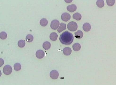
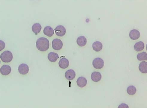
Figure 1. Blood smear from a cat infected with Myco - Figure 2. Blood smear from a cat infected with Myco - plasma organisms (Diff-Quick, magnification 1000 x). plasma organisms (Diff-Quick, magnification 1000x). Several Mycoplasma organisms are attached in an epi- Howell-Jolly bodies (black arrow), nuclear remnants, cellular location on erythrocytes (white arrows). A nu- should not be confused with red blood cell parasites. cleated red blood cell or normoblast (black arrow) is also Epicellular Mycoplasma organisms are indicated by present. white arrows.
duration. The cat was mainly kept indoors and was cur-
PCR, and revealed the presence of both Mycoplasma
rently on vaccinations. There was no history of recent
haemofelis (organism load of 2.4 x 10⁶/mL of blood)
drug administration or ectoparasite control. Recently,
and ‘Candidatus Mycoplasma haemominutum’ (or-
a young stray cat has also been adopted in the house-
ganism load of 1.5 x 10⁶/mL of blood).
hold. Physical examination revealed a lethargic cat
The cat tested negative for feline leukemia virus
with pale and mildly icteric mucous membranes, mild
(FeLV) antigen (ELISA) and feline immunodeficiency
tachycardia (180 beats/minute) and a low-grade sys-
virus (FIV) antibodies (immunofluorescence antibody
tolic murmur. Femoral pulses were bounding. Respi-
test). Lateral and ventrodorsal chest radiographs were
ratory rate and rectal temperature were normal (36
within normal limits. Abdominal ultrasound revealed
breaths/minute and 38.6°C, respectively). The right
mild diffuse splenomegaly with normal echogenicity.
prescapular lymph node was mildly enlarged. Ab-
Fine needle aspirates of the right prescapular lymph
node had a normal cellular distribution with the pre-
A complete blood count (CBC) revealed a severe
normochromic (mean corpuscular hemoglobin con-
Intravenous fluid therapy (Hartmann®) was given at
centration / MCHC 36.0 g/dL [reference 30-36 g/dL]),
maintenance rate. A fresh blood transfusion was con-
macrocytic (mean corpuscular volume / MCV 62 fl
sidered but postponed until absolutely necessary, be-
[reference 37-55 fl]), regenerative anemia (hematocrit
cause of financial concerns of the owner. Despite the
8.1% [reference 24-45%] with an absolute reticulocyte
severe anemia, the clinical and cardiorespiratory pa-
count of 338000/µL [reference < 40000/µL]). Leuko-
rameters remained stable during the following days.
cytes were within normal limits. The biochemistry
Medical treatment included doxycycline (Vi-
profile showed normal total protein (7.5 g/L [reference
bramycine® 5 mg/kg twice daily) combined with an im-
5.4-8.2 g/L]) and albumin concentrations (3.6 g/L [ref-
munosuppressive dose of parenteral dexamethasone
erence 2.7-4.5 g/L]), as well as normal renal parame-
(Rapidexon® 0.4 mg/kg once daily) initially, and oral
ters, liver enzymes and electrolytes. However, the
prednisolone (Prednisolone® 1 mg/kg twice daily) af-
bilirubin concentration was increased (2.8 mg/dL [ref-
ter the cat regained his appetite. A repeat blood smear
erence 0.1-0.6 mg/dL]). Urinalysis was normal except
on the second day of hospitalization no longer re-
for bilirubinuria. A direct polyvalent Coombs’ test
vealed Mycoplasma organisms. Signs of regeneration
(Nordic, Tilburg, the Netherlands) was found strongly
became more prominent (polychromasia, anisocytosis
positive (titer 1:4096). A fresh blood smear was stained
together with the presence of many normoblasts). A
with Diff-Quick. A regenerative response was clearly
CBC on the 6th day revealed a rising hematocrit
demonstrated by the presence of polychromasia, aniso-
(22.9%), with a MCV of 76.6 fl, the latter indicating the
cytosis and a moderate number of normoblasts and
presence of larger sized reticulocytes and thus an on-
Howell-Jolly bodies (Figures 1 and 2). Heinz bodies
going regenerative response. At that time, the systolic
were absent. Several erythrocytes showed one or more
murmur was no longer audible, suggesting that it was
epicellular organisms resembling Mycoplasma spp.
most likely caused by altered blood viscosity during the
(Figures 1 and 2). An EDTA-blood sample was sent by
regular mail to a veterinary laboratory specialized in
The cat was sent home with oral doxycycline (Vi-
molecular diagnostics for confirmation, characteriza-
bramycine®) 5 mg/kg twice daily for three weeks and
tion and quantification of the organisms by real-time
tapering doses of oral prednisolone (Prednisolone®, 1
Vlaams Diergeneeskundig Tijdschrift, 2012, 81
mg/kg twice daily starting dose). Furthermore, a
result in exposure of hidden antigens or changes in ex-
monthly flea prevention with fipronil (Frontline®) was
isting antigens, thereby activating the production of
prescribed. Four weeks later (and five days after dis-
host anti-erythrocyte antibodies. The anemia mostly re-
continuation of prednisolone), the physical examina-
sults from extravascular erythrophagocytosis by
tion, blood work, fresh blood smear, Coombs’ test and
macrophages in the spleen, liver, lungs and bone mar-
PCR for both Mycoplasma organisms were repeated.
row (Harvey, 2006). Other conditions, such as inflam-
According to the owner, the cat made full recovery and
matory, infectious and neoplastic diseases, may result in
was again alert and playful. The mucous membranes
the immune-mediated destruction of erythrocytes in cir-
were pink and the cardiac auscultation was normal, as
culation or lymphoid organs. A thorough work-up, in-
was the rest of the physical examination. The hemat-
cluding retroviral testing, chest radiographs and ab-
ocrit and red cell indices had returned within reference
dominal ultrasound, revealed no significant
limits (hematocrit 30%, MCV 45 fl, MCHC 36 g/dL).
abnormalities in the present case. The mild uniform
The bilirubin level was normalized (0,2 mg/dL) and
splenomegaly at initial presentation was believed to re-
bilirubinuria was no longer present. Other parameters
sult from sequestration of parasitized erythrocytes or
were still within normal limits. The blood smear re-
from extramedullary hematopoiesis. However, no fur-
vealed normal red cell morphology and the absence of
ther tests (fine-needle aspiration) were performed to
visible epicellular organisms. The Coombs’ test re-
sulted in a titer of 1:32. Real-time PCR revealed the
There is still debate on the exact natural route of
persistence of Mycoplasma haemofelis (hemoplasma
transmission of Mycoplasma spp. A role of Cteno-
load of 3.6 x 10⁵/mL of blood) and ‘Candidatus My-
cephalides felis has been proposed but is still unclear.
coplasma haemominutum’ (hemoplasma load of 3.2 x
Recently, DNA of Mycoplasma haemofelis and ‘Can-
106/mL of blood). Another three weeks of doxycy-
didatus Mycoplasma haemominutum’ has been am-
cline were advised but declined by the owner because
plified from blood of infected cats and from fleas col-
the cat had been clinically healthy since discontinua-
lected from client-owned cats by PCR (Lappin et al.,
tion and remained so during the following months.
2006). Woods et al. (2005) assessed the ability of fleasto transmit Mycoplasma spp. through hematophageous
activity and found that both species were ingested byC. felis when allowed to feed on experimentally in-
Anemia is a commonly encountered laboratory ab-
fected cats. Only Mycoplasma haemofelis was found to
normality in cats. It can be broadly classified into (1)
be transmitted from C. felis to cats by hematogenous
reduced hematopoiesis, such as anemia of inflamma-
route. However, the efficiency of infection was poor
tory disease, most FeLV infections and due to chronic
and clinical or hematological signs of anemia did not
renal failure, (2) external blood loss due to ectopara-
develop. Another study failed to detect the transmission
sites, tumors and hemostatic defects and (3) hemolysis.
of Mycoplasma spp. by oral ingestion of infected fleas
In the present case, the macrocytic regenerative ane-
or flea by-products (Woods et al., 2006). Recent stud-
mia, supportive of the increased presence of larger
ies also failed to show the successful transmission of
sized reticulocytes in the peripheral circulation), to-
‘Candidatus Mycoplasma turicensis’ infection between
gether with the normal serum protein concentration,
cats by the oral or subcutaneous inoculation of ‘Can-
hyperbilirubinemia and bilirubinuria, which is always
didatus Mycoplasma turicensis’-infected saliva
an abnormal finding in cats, were indicative of hemo -
(Museux et al., 2009). Aggressive cat interaction could
however result in transmission if a recipient cat is ex-
Hemolytic anemia in this species is mostly caused by
posed to blood of an infected cat (Museux et al., 2009).
acquired disorders, such as hemobartonellosis, feline in-
Despite the fact that many questions remain unans -
fectious peritonitis (FIP), and other infections (e.g.
wered, most reports as well as the present case support
FeLV subtype A), hypophosphatemia, oxidative agents
the recommendation to maintain flea control.
(e.g. onions, zinc and drugs) and primary and secondary
In the present case, it was not possible to determine
immune-mediated processes (Adams et al., 1993;
which of the two organisms was responsible for the
Robertson et al., 1998; Sykes, 2003; Norris et al., 2005;
clinical signs. Experimental infection with the large
Kohn et al., 2006). Less often, hemolysis is a conse-
variant, Mycoplasma haemofelis, resulted in more se-
quence of certain hereditary defects (Giger, 2005).
vere clinical signs than with the small variant, ‘Can-
Immune-mediated hemolytic anemia is considered
didatus Mycoplasma haemominutum’ (Westfall et al.,
to occur relatively rarely in cats compared to dogs, and
2001). The same observations were made in client-
is more often associated with secondary causes (Giger,
owned cats with naturally occurring hemobartonel-
2005; Kohn et al., 2006). In the present case, the direct
losis (Jensen et al., 2001). In the study of Westfall et al.
Coombs’ test was positive, indicating the presence of
(2001), cats with dual infections had the most severe
erythrocyte-bound antibodies and thus an immune-
clinical abnormalities. Proposed explanations included
mediated component to the hemolysis. Usually, most
dose-dependent effects or predisposition to immune-
damage to the red blood cell is not directly caused by
mediated disease in ‘Candidatus Mycoplasma
the parasite but rather because of immune-mediated in-
haemominutum’-infected cats that were subsequently
jury, especially in the acute phase of infection. The at-
exposed to the more pathogenic Mycoplasma haemofe-
tachment of the organisms to erythrocytes is thought to
Vlaams Diergeneeskundig Tijdschrift, 2012, 81
In the present case, the clinical course was rapid,
rofloxacin or marbofloxacin) would have resulted in
with only a two-day history of clinical signs before ad-
decreasing numbers of detectable microorganisms
mission with severe anemia. Another (retrospective)
(Dowers et al., 2002; Ishak et al., 2008). Cats may be-
study suggested that ‘Candidatus Mycoplasma
come PCR-positive again once the antibiotic treatment
haemominutum’ might be a primary pathogen in some
is stopped, or they can remain PCR-positive for months
naturally infected, immunocompetent cats (Reynolds
or years following infection, especially in the case of
and Lappin, 2007). Concurrent diseases may predis-
‘Candidatus Mycoplasma haemominutum’ infection
pose cats to develop more severe signs of anemia, as
(Tasker, 2006). Complete recovery with negative PCR
for example the presence of lymphoma (de Lorimier
results has recently been described in a Mycoplasma
and Messick, 2004). Pre-existing FeLV infection or
haemofelis infected cat, after a 6-week course of doxy-
FeLV-FIV co-infection also seems to potentiate the
cycline (Braddock et al., 2004). Alternatively, in a
severity of anemia caused by Mycoplasma haemofelis
study by Dowers et al. (2009), treatment with prad-
(George et al., 2002). However, the cat in this report
ofloxacin resulted in a more effective clearance of or-
tested negative for both retroviruses.
ganisms than with doxycycline. Another hypothesis for
The diagnosis is based on the detection of the or-
the persistent high bacterial load may be the concurrent
ganism in an epicellular location on feline erythro-
use of prednisolone, which might cause immunosup-
cytes on a fresh blood smear. However, this technique
pression with subsequent persistence of the organism.
has low sensitivity, especially in chronically infected
However, in a study by Dowers et al. (2002), treatment
animals, in cats with low parasite burden, or because
with immunosuppressive doses of methylprednisolone
of the cyclical parasitemia (Harvey, 2006; Tasker,
acetate did not result in the recurrence of organisms in
2006). Organisms rapidly detach from red blood cells
three cats after the successful treatment with doxycy-
in vitro, probably reflecting organism death a few
hours after blood collection (Tasker, 2006). Specificity
In this case, immunosuppressive doses of corticos-
may also be low, as organisms may be confused with
teroids were added to the treatment protocol because
Howell-Jolly bodies or staining artefacts, and it is not
the positive Coombs’ test strongly suggested an im-
possible to distinguish the two forms morphologically
mune-mediated component to the hemolysis (Harvey,
based on light microscopy alone (Jensen et al., 2001)
2006). This may indicate that one or both Mycoplasma
(Figure 2). Molecular techniques have greatly im-
species may have acted as haptens and triggered an im-
proved the ability to diagnose infections. The use of
mune-mediated destruction of affected erythrocytes.
real-time PCR offers several advantages over conven-
In conclusion, this case documents that cats in Bel-
tional PCR because it may both diagnose Mycoplasma
gium may be co-infected with Mycoplasma haemofe-spp. and quantify infectious burden (Tasker et al.,
lis and ‘Candidatus Mycoplasma haemominutum’,
2003). This is important for determining the signifi-
and that these organisms should be considered as pos-
cance of a positive PCR result in infections with My-coplasma haemofelis and ‘Candidatus Mycoplasmahaemominutum’, the latter not always resulting in clin-
ical disease in the host (Tasker et al., 2003). Further-more, quantification can help monitor the response to
Adams L.G., Hardy R.M., Weiss D.J., Bartges J.W. (1993).
treatment (Tasker et al., 2003). On the other hand, a
Hypophosphatemia and haemolytic anemia associated
positive cytology or PCR does not necessarily correlate
with diabetes mellitus and hepatic lipidosis in cats. Jour-
with clinical illness, as clinically normal cats can have
nal of Veterinary Internal Medicine 7, 266-271.
positive test results (Jensen et al., 2001). However,
Braddock J.A., Tasker S., Malik R. (2004). The use of real-
the more pathogenic form (Mycoplasma haemofelis) is
time PCR in the diagnosis and monitoring of Mycoplasma
rarely detected in cats without anemia (Jensen et al.,
haemofelis copy number in a naturally infected cat. Jour-nal of Feline Medicine and Surgery 6, 161-165.
de Lorimier L.-P., Messick J.B. (2004). Anemia associated
In the present case, the combination of doxycycline
with ‘Candidatus Mycoplasma haemominutum’ in a feline
and prednisolone resulted in rapid clinical and hemato-
leukemia virus-negative cat with lymphoma. Journal of
logical recovery, despite the lack of clearance of mi-
the American Animal Hospital Association 40, 423-427.
croorganisms as shown by the follow-up real-time PCR.
Dowers K.L., Olver C, Radecki S.V., Lappin M.R. (2002).
Total clearance of organisms, as confirmed by PCR,
Use of enrofloxacin for treatment of large-form Haemo-
seems to be rare, and most animals remain carriers
bartonella felis in experimentally infected cats. Journal of
(Dowers et al., 2002). Despite the persistence of both or-
the American Veterinary Medical Association 2, 250-253.
ganisms after antibiotic treatment in the present case, a
Dowers K.L., Tasker S., Radecki S.V., Lappin M.R. (2009).
tenfold decrease in Mycoplasma haemofelis load was
Use of pradofloxacin to treat experimentally induced My-
found by RT-PCR, and there was no longer evidence of
coplasma hemofelis infection in cats. American Journal of
ongoing hemolysis (normal hematocrit, MCV and biliru-
Veterinary Research 70, 105-111.
bin level). This may indicate that Myco plasma haemofe-
Duin M.B., Moyaert H., Pasmans F., Boyen F. (2009). He-
lis was responsible for the hemolytic anemia. After
motrofe mycoplasmen bij katten. Deel 1: literatuurover-
treatment, only the Coombs’ test was still borderline
zicht. Vlaams Diergeneeskundig Tijdschrift 78, 143-154.
positive. The question remains whether longer courses
Foley J.E., Pedersen N.C. (2001). ‘Candidatus Mycoplasma
haemominutum’, a low-virulence epierythrocytic para-
of doxycycline or an alternative antibiotic (e.g. en-
Vlaams Diergeneeskundig Tijdschrift, 2012, 81
site of cats. International Journal of Systematic and Evo-Journal of the American Animal Hospital Association 43,
lutionary Microbiology 51, 815-817.
Fujihara M., Watanabe M., Yamada T., Harasawa R. (2007).
Robertson J.E., Christopher M.M., Rogers Q.R. (1998). Heinz
Occurrence of ‘Candidatus Mycoplasma turicensis’ in-
body formation in cats fed baby food containing onion
fection in domestic cats in Japan. Journal of Veterinary
powder. Journal of the American Veterinary Medical As-Medical Science 69, 1061-1063.
Gentilini F., Novacco M., Turba M.E., Willi B., Bacci M.L.,
Sykes J.E. (2003). Feline hemotropic mycoplasmosis (feline
Hofmann-Lehmann R. (2009). Use of combined conven-
hemobartonellosis). Veterinary Clinics of North America:
tional and real-time PCR to determine the epidemiology
Small Animal Practice 33, 773-789.
of feline haemoplasma infections in northern Italy. Jour-
Tasker S., Helps C.R., Day M.J., Gruffydd-Jones T.J., Harbour
nal of Feline Medicine and Surgery 11, 277-285.
D.A. (2003). Use of Real-Time PCR to detect and quantify
George J.W., Rideout B.A., Griffey S.M., Pedersen N.C. Mycoplasma haemofelis and ‘Candidatus Mycoplasma
(2002). Effect of preexisting FeLV or FeLV and feline im-
haemominutum’ DNA. Journal of Clinical Microbiology
munodeficiency virus coinfection on pathogenicity of the
small variant of Haemobartonella felis in cats. American
Tasker S. (2006). Current concepts in feline haemobar-
Journal of Veterinary Research 63, 1172-1178.
tonellosis. Companion Animal Practice 28, 136-141.
Giger U. (2005). Regenerative anemias caused by blood
Westfall D.S., Jensen W.A., Reagan W.J., Radecki S.V.,
loss or hemolysis. In: Ettinger S.J., Feldman E.C. (editors).
Lappin M.R. (2001). Inoculation of two genotypes of He-Textbook of Veterinary Internal Medicine – Diseases of themobartonella felis (California and Ohio variants) to induce
Dog and Cat. 6th Edition, Elsevier Saunders, Philadel-
infection in cats and the response to treatment with
azithromycin. American Journal of Veterinary Research
Harvey J.W. (2006). Hemotrophic Mycoplasmosis (Hemo-
bartonellosis). In: Greene C.E. (editor). Infectious Dis-
Willi B., Boretti F.S., Cattori V., Tasker S., Meli M.L.,
eases of the Dog and Cat. 3rd Edition. Elsevier Saunders,
Reusch C., Lutz H., Hofmann-Lehmann R. (2005). Iden-
tification, molecular characterization, and experimental
Ishak A.M., Dowers K.L., Cavanaugh M.T., Powell C.C.,
transmission of a new hemoplasma isolate from a cat with
Hawley J.R., Radecki S.V., Lappin M.R. (2008). Mar-
hemolytic anemia in Switzerland. Journal of Clinical Mi-
bofloxacin for the treatment of experimentally induced
Mycoplasma haemofelis infection in cats. Journal of Vet-
Willi B., Tasker S. Boretti F.S., Doherr M.G., Cattori V., Meli
erinary Internal Medicine 22, 288-292.
M.L., Lobetti R.G., Malik R., Reush C.E., Lutz H., Hof-
Jensen W.A., Lappin M.R., Kamkar S., Reagan W.J. (2001).
mann-Lehmann R. (2006). Phylogenetic analysis of ‘Can-
Use of a polymerase chain reaction assay to detect and dif-
didatus Mycoplasma turicensis’ isolates from pet cats in
ferentiate two strains of Haemobartonella felis in naturally
the United Kingdom, Australia, and South Africa, with
infected cats. American Journal of Veterinary Research 62,
analysis of risk factors for infection. Journal of Clinical
Kohn B., Weingart C., Eckmann V., Ottenjann M., Leibold
Woods J.E., Brewer M.M., Hawley J.R., Wisnewski N.,
W. (2006). Primary immune-mediated hemolytic anemia
Lappin M.R. (2005). Evaluation of experimental trans-
in 19 cats. Journal of Veterinary Internal Medicine 20,
mission of ‘Candidatus Mycoplasma haemominutum’ and
Mycoplasma haemofelis by Ctenocephalides felis to cats.
Lappin M.R., Griffin B., Brunt J., Riley A., Burney D., Haw-
American Journal of Veterinary Research 66, 1008-1012.
ley J., Brewer M.M., Jensen W.A. (2006). Prevalence of
Woods J.E., Wisnewski N., Lappin M.R. (2006). Attempted
Bartonella species, haemoplasma species, Ehrlichia species,
transmission of ‘Candidatus Mycoplasma haemominu-
Anaplasma phagocytophilum, and Neorickettsia risticii
tum’ and Mycoplasma haemofelis by feeding cats infected
DNA in the blood of cats and their fleas in the United
Ctenocephalides felis. American Journal of Veterinary
States. Journal of Feline Medicine and Surgery 8, 85- 90.
Museux K., Boretti F.S., Willi B., Riond B., Hoelzle L.E.,
Wittenbrink M.M., Tasker S., Wengi N., Reusch C.E.,Lutz H., Hofmann-Lehmann R. (2009). In vivo transmis-sion studies of ‘Candidatus Mycoplasma turicensis’ in adomestic cat. Veterinary Research 40, 45.
Neimark H., Johansson K.E., Rikihisa Y, Tully J.G. (2001).
Proposal to transfer some members of the genera Haemo-bartonella and Eperythrozoon to the genus Mycoplasmawith descriptions of ‘Candidatus Mycoplasma haemofe-lis’, ‘Candidatus Mycoplasma haemomuris’, ‘CandidatusMycoplasma haemosuis’ and ‘Candidatus Mycoplasmawenyonii. International Journal of Systematic and Evo-lutionary Microbiology 51, 891-899.
Norris J.M., Bosward K.L., White J.D., Baral R.M., Catt
M.J., Malik R. (2005). Clinicopathological findings asso-ciated with feline infectious peritonitis in Sydney, Aus-tralia: 42 cases (1990-2002). Australian Veterinary Jour-nal 83, 666- 673.
Reynolds C.A., Lappin M.R. (2007). ‘Candidatus Myco -
plasma haemominutum’ infections in 21 client-owned cats.
Richmond upon Thames LGBT Forum Appendix 3 - Drug Misuse and the LGBT Community Submission to the Richmond upon Thames Drug Misuse Scrutiny Task Group – 31 January 2011 Introduction The Richmond upon Thames Lesbian, Gay, Bisexual and Trans (LGBT) Forum is a voluntary community group providing a voice for LGBT people who live, work, study or visit in the borough. We were
4Life Transfer Factor® GluCoach ™ EQUILIBRE A SUA BOA SAÚDE Manter níveis saudáveis de glucose (açúcares) é uma parte importante/significativa da saúdepancreática e ainda mais um aspecto importante da boa saúde de milhões de pessoas do nosso mundo. Desequilíbrios (do foro) metabólicos e endócrinos impede o nosso organismo de produzir ou de utilizarpropriamente insulina, uma h
 Vlaams Diergeneeskundig Tijdschrift, 2012, 81
Figure 1. Blood smear from a cat infected with Myco -
Vlaams Diergeneeskundig Tijdschrift, 2012, 81
Figure 1. Blood smear from a cat infected with Myco -