James C. Wittig, MD Orthopedic Oncologist Percutaneous Radiofrequency Ablation (RFA)
The latest, “State of the Art Treatment” for Osteoid Osteoma is PERCUTANEOUS RADIOFREQUENCY ABLATION (also known as RFA). This is a minimally invasive procedure that is performed under a CAT Scan, usually by a highly specialized musculoskeletal radiologist, in which a needle or probe is inserted into the lesion and the lesion is heated and destroyed. The CAT scan is utilized to localize the Osteoid Osteoma so the needle can be guided directly into the tumor. It is an outpatient procedure. The patient goes home the same day. It is minimally invasive and therefore only a small stab incision or poke hole is made for the needle. The procedure does require that the patient be put to sleep with general anesthesia because insertion of the needle into the Osteoid Osteoma is very painful. The patient must also lie motionless during the procedure. The procedure is greater than 90% effective. This is the same success rate as with actual surgical removal. The pain from the osteoid osteoma is usually relieved within 1 day. Often in the recovery room after the procedure, the patient will say that the pain from the tumor is gone. There is full use of the leg or arm and return to normal activities the following day. There is virtually no blood loss and very little risk (less than 1% risk) of developing an infection after the procedure. Less than 10% of the time the procedure needs to be repeated or the patient requires a surgical procedure to remove the tumor.
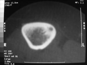
These are the CT scans of a patient who underwent RF Ablation of an osteoid osteoma of the tibia. The first CT scan demonstrates the nidus (arrow) of the osteoid osteoma. The second CT scan demonstrates the tiny RF ablation probe (arrow) being inserted into the nidus through a small needle.
This is a photograph of the procedure being performed. The RF ablation probe (Blue Arrow) is inserted through a needle into the nidus. The needle (Yellow arrow) is utilized to localize the nidus in the CT scan and drill a hole in the bone to allow the RF ablation probe to be inserted into the nidus. Notice the procedure is minimally invasive.
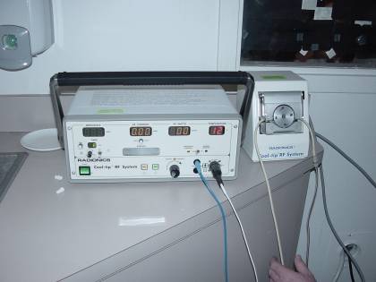
The RF ablation probe is attached to the generator which heats and destroys the osteoid osteoma.
Other treatment alternatives include surgical excision or simply treating the pain with chronic, non-steroidal anti-inflammatory medication like aspirin, naprosyn, ibuprofen or motrin. Surgical excision of the lesion has always been the traditional treatment for an Osteoid Osteoma. Due to the extremely small size of an Osteoid Osteoma, it can be very difficult to identify the tumor during surgery. It is like “LOOKING FOR A NEEDLE IN A HAYSTACK”. The area of the bone containing the Osteoid Osteoma would be localized during surgery. A large piece of bone would need to be removed to ensure the Osteoid Osteoma was removed with the bone. Osteoid Osteomas are so small; they can be extremely difficult to localize accurately during surgery. Therefore, a large piece of bone in the proposed area of the tumor is often removed. A large incision is required. It is obvious that this type of surgery is much more extensive and associated with many more potential problems or complications compared to RF ABLATION. This type of surgery often requires the bone to be protected by placing a metal rod inside the bone or by fastening a plate on the bone with screws to prevent the bone from fracturing or breaking after the surgery. Additionally, a bone grafting procedure may need to be performed to fill in the defect in the bone There is also much higher risk of infection, blood loss, nerve dysfunction and scar tissue formation. There is more pain and the patient usually remains in the hospital for several days. A prolonged recovery period as well as physical therapy and rehabilitation are required. The surgical pain may last for months, requiring narcotic pain medications. If the tumor is capable of being localized accurately during surgery (much of this depends on surgical expertise), a small drill can often be utilized (Procedure referred to as “Burr- Down Resection”) to remove the tumor without removing a lot of bone. Sometimes, a needle can be placed into the tumor under a CT scan to localize the tumor for the surgeon. (similar to how the needle is inserted for RF ABLATION) The patient is then transported to the operating room where the surgical procedure is performed. The needle helps the surgeon determine the exact location of the tumor so that “Burr Down Resection” with a dental type drill can be performed more easily and accurately. This is to prevent the tumor from being missed during the surgery. There are also many more potential complications with this procedure than with RF ABLATION. Although the procedure is less invasive than the surgical procedure described above, it is still associated with an incision, blood loss, higher risk of infection compared to RF ABLATION. It may also require fixation of the bone to prevent fracture (not as often as surgical procedure described above). It usually does
not require bone grafting. There is prolonged recuperation, hospital stay, postoperative pain and rehabilitation compared to RF ABLATION. An Osteoid Osteoma is not a malignant or cancerous tumor. It also does not usually grow more than 2 centimeters and therefore does not destroy the bone from which it arises. It is therefore not absolutely necessary to remove or invasively treat the tumor if pain medication works in relieving the pain. The only reason to treat the tumor is for pain relief. In this situation the tumor would be observed with periodic X-rays and CAT scans. The tumor may spontaneously disappear or resolve. This usually takes at least a few years. The patient would be treated with a non-steroidal anti-inflammatory medication like naprosyn, motrin, aspirin or ibuprofen for this time period. The pain often lasts for years. Some patients have reported pain for over 10 years. Treatment with pain medication is therefore usually for a prolonged period and patients usually require the pain medication for 24 hours a day. I have had patients who have been treated in this manner, including children, who have developed gastric ulcers and kidney failure. In addition, parents usually complain that despite the pain medication, their children still wake up at night and do not have a peaceful night of sleep. It is also difficult for parents to make sure that the child is getting the pain medication in school. Thus, when the pain medication wears off, the child is in pain. This causes difficulties with school and with interacting with other children. The sleeping difficulties further complicates their school related activities and learning. What are the risks associated with Radiofrequency Ablation?
Infection----less than 1% risk Persistent Pain or Failure to Eradicate the Tumor Completely: A second RF Ablation procedure or a surgical procedure may be required. Recurrence of the Osteoid Osteoma: The tumor may come back up to two years after the procedure. This is unusual (less than 10% chance) but the patient needs to be monitored after the procedure. The patient will begin to experience the pain again if the tumor comes back (recurs). Please note: there will always be a persistent scar in the bone that can be seen on an X-ray after the procedure. Skin Burn: This is a rare event however osteoid osteomas that are adjacent to the skin may predispose the skin to a small burn. Care is taken to ensure that the probe is buried within the nidus to minimize the risk of a burn. What do I need to do if I have been diagnosed with an Osteoid Osteoma and I want to have Radiofrequency Ablation performed?
You should set up an appointment to see Dr. James C. Wittig, Orthopedic Oncology, to ensure that you have had all the appropriate tests needed to diagnose the Osteoid Osteoma. You will need to be examined and all of the actual radiological studies will need to be reviewed by Dr. Wittig. If Dr. Wittig agrees with the diagnosis he will schedule you for the procedure with one of the radiologists whom he works closely with and who has experience with radiofrequency ablation. Dr. Wittig will need to keep your radiological studies to discuss them with the radiologist* * Special thanks to musculoskeletal radiologists Leon Rybak and George Nomikos for helping to develop this informational section on osteoid osteoma and radiofrequencey ablation.
Journal of Psychiatric Research 43 (2009) 1086–1094Contents lists available at ScienceDirectj o u r n a l h o m e p a g e : w w w . e l s e v i e r . c o m / l o c a t e / j p s y c h i r e sAssociation of trait-defined, eating-disorder sub-phenotypes with(biallelic and triallelic) 5HTTLPR variationsHoward Steiger *, Jodie Richardson, Norbert Schmitz, Ridha Joober, Mimi Israel, Kenneth
IN DEFENSE OF METAPHOR "That's how they all squeal at first," he said. "As if the world could be changed without killing someone."Friedrich Dürrenmatt, Grieche sucht Griechin Mephistopheles: No Lord, I believe that, as always,A few years ago, the papers announced that the government ofSouth Africa was going to set up a programme to import and producelow-cost drugs to trea
 James C. Wittig, MD
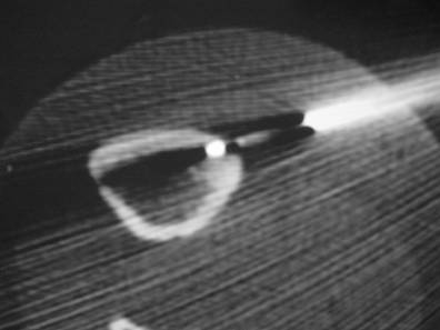
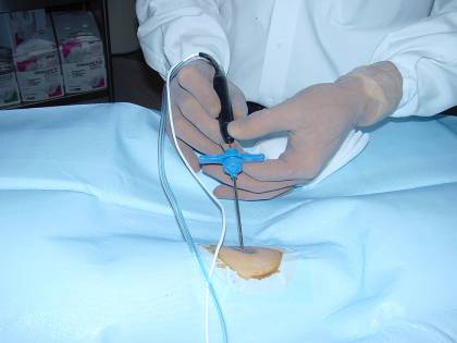 This is a photograph of the procedure being performed. The RF ablation probe (Blue Arrow) is inserted through a needle into the nidus. The needle (Yellow arrow) is utilized to localize the nidus in the CT scan and drill a hole in the bone to allow the RF ablation probe to be inserted into the nidus. Notice the procedure is minimally invasive.
The RF ablation probe is attached to the generator which heats and destroys the osteoid osteoma.
This is a photograph of the procedure being performed. The RF ablation probe (Blue Arrow) is inserted through a needle into the nidus. The needle (Yellow arrow) is utilized to localize the nidus in the CT scan and drill a hole in the bone to allow the RF ablation probe to be inserted into the nidus. Notice the procedure is minimally invasive.
The RF ablation probe is attached to the generator which heats and destroys the osteoid osteoma.