Article gastrointestinal disorders Gastroschisis: Embryology, Pathogenesis, Epidemiology Shilpi Chabra, MD,*
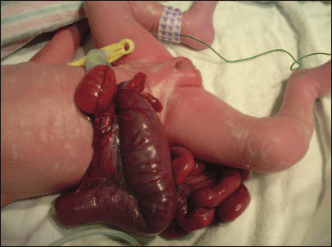
Objectives After completing this article, readers should be able to: 1. Describe normal embryology and various theories contributing to derangements in development leading to gastroschisis. 2. Delineate several theories regarding the pathogenesis of gastroschisis. 3. Explain the environmental and other risk factors linked to gastroschisis. 4. Describe the prevalence of gastroschisis in developed countries and various theories explaining it. Introduction Gastroschisis is a congenital anterior abdominal wall defect, adjacent and usually to the right of the umbilical cord insertion. It occurs as a small, full-thickness periumbilical cleft either immediately adjacent to the umbilicus or separated from it by a strip of skin. This results in herniation of the abdominal contents into the amniotic sac, usually just the small intestine, but sometimes also the stomach, colon, and ovaries (Figure). The abdominal wall defect is relatively small compared with the size of the eviscerated bowel, which often develops walls that are matted and thickened with a fibrous peel. Gastroschisis has no covering sac and no associated syndromes. This differentiates it from an omphalocele, which usually is covered by a membranous sac and more frequently is associated with other structural and chromosomal anomalies (Table 1). In addition, although gastroschisis may be associated with gastrointestinal anomalies such as intestinal atresia, stenosis, and malrotation, it has a much better prognosis than omphalocele. Historical Perspective The term gastroschisis is derived from the Greek word laproschisis, meaning “bellycleft.” It was used in the 19th and early 20th centuries by teratologists to designate all abdominal wall defects. No clear distinctions were made between abdominal wall defects until 1953, when Moore and Stokes classified them based on their appearance at birth. They suggested that the term gastroschisis be reserved for those cases in which the defect is adjacent to the normally inserted umbilical cord and there is no evidence of a sac covering the extruded viscera. Although the first report of a case of gastroschisis was in 1733, the first report of successful closure of a small abdominal wall defect was not until 1943 by Watkins, a surgeon from Virginia. Embryology The pathogenesis of gastroschisis remains controversial. To understand various theories regarding this defect, it is essen- Abbreviations
The gastrointestinal tract develops from the primitive
International Clearinghouse of Birth Defects
digestive tube derived from the yolk sac. Early in gestation, a
portion of the gut opens ventrally into the yolk sac—the
International Classification of Diseases,
midgut. At 31⁄2 weeks’ gestation, the gut becomes distinct
from the yolk sac. The embryonic disk is folded into cephalic,
ICD-9-CM: International Classification of Diseases,
caudal, and lateral folds, each of which converges at the
umbilicus to obliterate the coelom, which forms the future
*Assistant Professor of Pediatrics, University of Washington, Seattle, Wash. †Division Head and Professor of Pediatrics, Department of Pediatrics, University of Washington, Seattle, Wash. NeoReviews Vol.6 No.11 November 2005 e493 gastrointestinal disorders gastroschisis
that subsequently ruptures, resulting in visceral hernia-tion. The left omphalomesenteric artery also involutes,and the right one develops into the superior mesentericartery. Similarly, disruption of this process may causeischemia and the development of gastroschisis. Othertheories speculating on the embryologic origins of gas-troschisis are summarized in Table 2. These includerupture of an omphalocele and various intrauterine in-sults. Infarction due to a vascular insult and strangulationof the eviscerated bowel by the contracting umbilical ringor midgut volvulus are proposed in the pathogenesis ofintestinal atresia and other gastroschisis variants, whichare summarized in Table 3. However, the vascular theoryis supported by the association of gastroschisis with ma-ternal smoking and other anomalies also known to have a
Figure. Gastroschisis, resulting in herniation of the abdominal contents into the amniotic sac.
Animal models have been developed in chicks, rab-
bits, lambs, and mice and may hold the key to the origin
peritoneal cavity. At the beginning of the 6th week, the
of gastroschisis. Most of these models have used terato-
midgut elongates at a rate faster than that of the elonga-
gens that affect fetal vasculature (Table 4).
tion of the embryonic body. This results in physiologicdevelopment of an umbilical hernia. At 10 weeks, themidgut returns rapidly to the embryonic abdominal cav-
Pathogenesis
ity, and the layers of the cephalic, caudal, and lateral folds
Gastroschisis is an isolated structural anomaly, and no
join to close the defect in the abdominal wall. This
single definitive genetic or environmental cause has been
normal reduction of the physiologic midgut herniation
identified. However, numerous theories have been pro-
followed by abdominal wall closure is key to normal
development. Various theories have been developed toexplain how or why this does not happen.
The vascular disruption theory is held most com-
Gastroschisis is primarily an isolated defect occurring
monly. The embryo begins with two umbilical veins and
sporadically and having a multifactorial etiology. How-
two omphalomesenteric arteries. Between 28 and
ever, familial clusters and occurrence in twins suggest a
32 days after conception, the right umbilical vein invo-
role of heredity and an autosomal inheritance pattern
lutes. One gastroschisis theory is that premature involu-
with variable expression. Sibling recurrence rates range
tion can lead to ischemia, which results in a weak spot
from 3% to 5%, which emphasizes the need for appropri-
Table 1. Differences Between Gastroschisis and Omphalocele Gastroschisis Omphalocele Incidence 1 in 10,000 (now increasing) 1 in 5,000 Defect Location Right paraumbilical Covering Sac Present (unless sac ruptured) Description Free intestinal loops Firm mass including bowel, liver, etc Associated With Prematurity 50% to 60% 10% to 20% Necrotizing Enterocolitis Common (18%) Uncommon Common Associated Anomalies Gastrointestinal (10% to 25%) Trisomy syndromes (30%)
● Intestinal atresia Cardiac defects (20%)
● Malrotation Beckwith-Weidemann syndrome Cryptorchidism (31%) Bladder extrophy Prognosis Excellent for small defect Varies with associated anomalies Mortality Varies with associated anomalies (80% with cardiac defect)
e494 NeoReviews Vol.6 No.11 November 2005 gastrointestinal disorders gastroschisis Table 2. Theories Regarding Embryogenesis of Gastroschisis Duhamel (1963) Teratogenic insult resulting in defective differentiation of the somatopleural mesenchyme Shaw (1975) Rupture of a hernia of the umbilical cord at the site of involution of the right umbilical vein DeVries (1980) Abnormal right umbilical vein atrophy resulting in weakness and defect of abdominal wall, with failure of epidermal differentiation Van Allen (1981, 1987) Vascular disruption theory Hoyme (1981, 1983) Omphalomesenteric artery insult with disruption of umbilical ring
the risk of fetal gastroschisis 3.6-fold and the risk of small intestinalatresia 4.2-fold.
ate counseling for recurrence in a family that has a history
Young maternal age and primigravida status have
of gastroschisis. Experimental studies have attempted to
been associated with increasing prevalence of gastroschi-
identify a gene responsible for gastroschisis. In 1998, a
sis. However, young paternal age has not been identified
study in mice implicated the region of mouse chromosome
as a significant risk factor. The association of gastroschisis
7 in the pathogenesis of radiation-induced gastroschisis. In
with younger women who could have increased use of
another study, induction of mutation in the bone morpho-
illicit drugs, alcohol, and smoking may point to a poten-
genic protein-1 gene in mice resulted in a condition similar
tial factor associated with their lifestyle. Other factors
to gastroschisis; unfortunately, no mutation of this gene has
associated with increased risk are low pregnancy body
been found in affected human infants. This lack of evidence
mass index and maternal diet. The teenage diet may have
for genetic predisposition further emphasizes the need to
low levels of alpha-carotene and total glutathione and
identify possible environmental factors.
high levels of nitrosamines, which may suggest a patho-genetic role for a nutrient deficiency. Environmental
The trend of increasing birth prevalence of gastroschisis
Epidemiology
in different populations at different time periods over a
The prevalence of gastroschisis has increased internation-
wide geographic distribution suggests possible exposure
ally (Table 7). This increased prevalence of gastroschisis
to environmental teratogens (Table 5). Current research
(but not of omphalocele) was first observed in Finland in
has focused on vasoactive drugs. Epidemiologic studies
the 1970s. In the 1980s, increases were observed in
have shown an increased risk of gastroschisis in mothers
Strasbourg, Paris, Israel, and Atlanta, as reported by the
who have reported taking vasoactive over-the-counter
International Clearinghouse of Birth Defects Monitor-
medications, including pseudoephedrine, phenylpropa-
ing Systems (ICBDMS). Reports of increased prevalence
nolamine, aspirin, ibuprofen, and acetaminophen. Aspi-rin has been shown to increase the risk for gastroschisis in
Table 3. Gastroschisis Variants
both animal and human studies. In one mouse study,gastroschisis developed after aspirin administration on
Proposed Pathogenesis
day 9 of gestation (corresponds to the 4th week of
Left-sided Gastroschisis Regression of left umbilical
human gestation), but not when aspirin was adminis-
tered on days 10 and 12, implicating a window of devel-
Vanishing Gut Midgut volvulus and bowel
opmental vulnerability. Use of these types of medications
infarction
during upper respiratory tract infections suggests the
Closed Gastroschisis Tightening of abdominal
possibility of an underlying infectious agent as another
fascial defect around the bowel, causing ischemia,
potential etiologic factor. Various risk factors associated
resorption, and
with gastroschisis are summarized in Table 6. These
spontaneous closure
factors cover a wide spectrum, including parental occu-
NeoReviews Vol.6 No.11 November 2005 e495 gastrointestinal disorders gastroschisis Table 4. Animal Models of Gastroschisis Investigator (Year)
10,000 total births, and there wasno significant overall linear trend
Kimmel (1971) Salicylates Nielsen (1986) Hyperthermia Randell (1994) Hillebrandt (1998) Irradiation during preimplantation Singh (2003) Protein and zinc deficiency with carbon monoxide exposure
periods, with a low, stable rate ofgastroschisis from 1968 through1975 (0.8 per 10,000 births) and a
of gastroschisis continued into the 1990s, along with
higher, stable rate from 1976 through 2000 (2.3 per
geographic variations attributed to maternal age. Around
10,000 births), and no temporal trend observed since
the same time period, reports of an increased prevalence
of gastroschisis in England showed geographic variationwithin the country. Several of these epidemiologic stud-
Maternal Age
ies have demonstrated an increased prevalence of gastros-
Maternal age of less than 20 years has been identified as a
significant risk factor for gastroschisis, especially in devel-
In the United States, a similarly increased prevalence
oped countries. The reasons for this association remain
of gastroschisis has been reported in several states, which
unclear, although speculations of an unidentified terato-
varies widely geographically (Table 8). In the past, the
gen related to modern lifestyle factors, including hob-
ratio of the number of cases of omphalocele to gastro-
bies, occupation, and diet, have been made. An associa-
schisis has been reported to be 3:2. However, some have
tion with nulliparity and the use of oral contraceptives
suggested that the prevalence of gastroschisis has been
during conception could point toward teratogenic
increasing in contrast to a stable or decreasing prevalence
of omphalocele, thus altering the ratio. In Florida, forexample, a study analyzing cases of abdominal wall de-
Maternal Race
fects between 1982 and 1999 reported the ratio of
Gastroschisis has been reported with increased frequency
number of cases of omphalocele to gastroschisis to be
in Hispanics. In a study from Utah, 23% of the gastros-
chisis cohort was Hispanic. A retrospective study from
This increased prevalence occurs in phases over differ-
Mexico reported one of the highest prevalence rates of
ent time periods. For example, a study in Denmark that
gastroschisis of 4.93 per 10,000 in 1998 among persons
evaluated abdominal wall defects data from 20 birthcohorts in three nationwide registries showed an initialphase of increase in gastroschisis until 1976, followed by
Table 6. Risk Factors Associated
a decrease to the initial 1970s value in 1983, and asubsequent new increase. The average point prevalence
With Gastroschisis
● Parental occupation (eg, printer/computer manufacturing factories) Table 5. Potential Teratogens
● Young maternal age ● Hispanic race Associated with Gastroschisis
● Poor maternal education ● Low socioeconomic status
● Organic chemicals/solvents
● Lack of prenatal care
● Cyclooxygenase inhibitors (aspirin, ibuprofen)
● Nulliparity
● Decongestants
● More than one elective abortion
● Acetaminophen
● Short interval between menarche and first pregnancy
● Oral contraceptives
● Chorionic villus sampling
● Maternal smoking
● Residence surrounding landfill sites
● Alcohol
● Maternal diet (low alpha-carotene, low total
● Illicit drugs (eg, cocaine, amphetamine) glutathione, high nitrosamines)
● X-ray irradiation in early pregnancy
● Low pregnancy body mass index
e496 NeoReviews Vol.6 No.11 November 2005 gastrointestinal disorders gastroschisis Geographic Distribution Table 7. Increased Prevalence of
An association of gastroschisis with a behavioral or envi-
Gastroschisis in Developed
ronmental exposure has been postulated due to its asso-ciation with geographic variations in different parts of the
Countries
world. The prevalence of gastroschisis may vary between
Incidence per
rural and urban regions, although information in the
10,000 Births
literature is insufficient. In a Finnish study, an apparent
1970 to 1974
increase in prevalence of gastroschisis was noted in
1975 to 1979
northern Finland only, and urban residence was a corre-
England and Wales
late of the increased gastroschisis risk. In the United
States, there have been reports of disparity in distribution
Southwestern England
of gastroschisis, such as preponderance in the rural part
Northern England
of New York. A retrospective study of a cluster of gas-
troschisis from Kentucky found no evidence of temporal
or spatial clustering in the cases, and there was no asso-
1996 to 1997
ciation with county of maternal residence. Anomalies of
Western Australia 1980 to 1990
the central nervous system, orofacial clefts, and limb
1985 to 1990
reduction defects have been associated with parental
1996 to 2000
exposure to pesticides. The risk of gastroschisis in rural
areas, especially with farming communities, could be
increased due to use of pesticides or fertilizers, but no
1967 to 1974
definitive studies have confirmed this hypothesis. 1995 to 1998 Seasonal Association
Conflicting reports have suggested an association of gas-
*International Clearinghouse for Birth Defects Monitoring Systems
troschisis with month of birth, raising the question of aninfectious cause. One such study reported that 37% ofgastroschisis conceptions occurred during the first quar-
of Mexican origin. A study from Hawaii found a de-
ter of the year. In another study, infants born during
creased prevalence in Far East Asians. In New York, there
January, February, or March were at greater risk. How-
was a higher mortality rate for black infants who had
ever, yet another study found that month of birth was not
gastroschisis compared with whites, and a recent study in
associated with gastroschisis. Due to the seasonal varia-
Atlanta found that the infants who had gastroschisis born
tions reported and association with medications used for
to teenage mothers and mothers 20 to 24 years of age
respiratory illnesses, a viral cause is still speculated.
were less likely to be born to black mothers than to whitemothers. Other Factors
Although the recent rapid increases in the rate of gastros-chisis have been linked primarily to environmental fac-
Table 8. Increased Prevalence of
tors, several other theories have been offered. For exam-ple, lack of differentiation between the clinical forms of
Gastroschisis in the United States
abdominal wall defects either due to selection bias, pre-vious underreporting of the defect, or misclassification of
Incidence per 10,000 Births
gastroschisis as omphalocele could be a factor. In aSwedish study, 8% of cases of abdominal wall defects
were “unclassifiable,” and in a Danish study, 20% of cases
of gastroschisis were misclassified. In British Columbia,
there were no reports of gastroschisis before 1969 be-
North Carolina
cause the condition was unknown, and previous cases
were diagnosed as omphalocele. Another theory regard-
Atlanta, Georgia 1968 to 1975
ing the increased prevalence of gastroschisis suggests that
1976 to 2000
shifts in maternal age distribution and selected termina-
NeoReviews Vol.6 No.11 November 2005 e497 gastrointestinal disorders gastroschisis
tion in fetuses have occurred due to improvements in
smoking, leading to speculations of a teratogen related to
prenatal diagnosis. A study from the ICBDMS postu-
modern lifestyle that remains to be identified. Also, it is
lated underreporting of cases in countries such as France
possible that gastroschisis may be related to a combina-
and Netherlands, where there is a high proportion of
tion of factors working synergistically, rather than an
selective terminations. This also was suggested in a Dan-
isolated single event or exposure. This rising prevalence
ish study after induced abortions were legalized in Den-
of gastroschisis has been described as an epidemic, em-
mark in 1973. One study reported the incidence of
phasizing the importance of continued monitoring and
abdominal wall defects in stillborn infants to be 20 times
evaluation of pathogenetic factors. The potential associ-
ation of gastroschisis with medications, diet, and other
One of the disadvantages of the epidemiologic studies
maternal factors could have implications for pregnancy
evaluating changes in prevalence is the reliance on vari-
planning similar to neural tube defects. Thus, it is an
able reporting of data to birth defects surveillance pro-
important public health issue, highlighting the need for a
grams. For example, some studies include elective termi-
more complete multicenter epidemiologic study.
nations in their calculations, thereby increasing theirreported occurrence. Several retrospective studies useICD-9 codes to identify patients. However, it is impor-tant to clarify that the code for gastroschisis is the same as
Suggested Reading
for omphalocele. In fact, the ICD-9 code includes all
Baerg J, Kaban G, Tonita J, Pahwa P, Reid D. Gastroschisis:
congenital anomalies of the abdominal wall (756.79
A sixteen-year review. J Pediatr Surg. 2003;38:771–74
Calzolari E, Bianchi F, Dolk H, Milan M. Omphalocele and gas-
Anomalies of the abdominal wall, other congenital
troschisis in Europe: a survey of 3 million births 1980 –1990.
anomalies of abdominal wall). Currently, individual de-
EUROCAT Working Group. Am J Med Genet. 1995;58:
fects cannot be separately identified based on ICD-9
codes, and chart review and medical record data are
Forrester MB, Merz RD. Epidemiology of abdominal wall defects,
needed to separate abdominal wall defects accurately.
Hawaii, 1986 –1997. Teratology. 1999;60:117–123
Perhaps, the time is right to petition for a new ICD-9
Goldbaum G, Daling J, Milham S. Risk factors for gastroschisis.
code or make clinical modifications to it, identified as
Hwang PJ, Kousseff BG. Omphalocele and gastroschisis: an 18-year
ICD-9-CM, in an attempt to separate these vastly differ-
review study. Genet Med. 2004;6:232–236
ent anomalies and improve data collection for future
Laughon M, Meyer R, Bose C, et al. Rising birth prevalence of
gastroschisis. J Perinatol. 2003;23:291–293
Nichols CR, Dickinson JE, Pemberton PJ. Rising incidence of
gastroschisis in teenage pregnancies. J Matern Fetal Med. 1997;6:225–229
Epidemiologic studies from the United States and other
Torfs CP, Velie EM, Oechsli FW, Bateson TF, Curry CJ. A popu-
developed countries around the globe have reported an
lation-based study of gastroschisis: demographic, pregnancy,
increased prevalence of gastroschisis over a wide geo-
and lifestyle risk factors. Teratology. 1994;50:44 –53
graphic distribution. Although environmental and ma-
Werler MM, Sheehan JE, Mitchell AA. Association of vasoconstric-
ternal factors have been suspected, the cause of gastros-
tive exposures with risks of gastroschisis and small intestinalatresia. Epidemiology. 2003;14:349 –354
chisis remains unclear, and no single cause has yet been
Werler MM, Sheehan JE, Mitchell AA. Maternal medication use
implicated. Universally, there is a significant association
and risks of gastroschisis and small intestinal atresia. Am J
of gastroschisis with young maternal age along with
e498 NeoReviews Vol.6 No.11 November 2005 gastrointestinal disorders gastroschisis NeoReviews Quiz 1. Gastroschisis is a congenital anterior abdominal wall defect, adjacent and usually to the right of the umbilical cord insertion. Of the following, the most common anomaly associated with gastroschisis is: A. Beckwith-Wiedemann syndrome. B. Congenital heart defect. C. Cryptorchidism. D. Trisomy 21. E. Urinary bladder exstrophy. 2. The pathogenesis of gastroschisis remains controversial, although several theories have been proposed to explain its development. Of the following, the most commonly held theory of the pathogenesis of gastroschisis is: A. Ethanol exposure during early embryogenesis. B. Irradiation during preimplantation. C. Protein and zinc deficiency with carbon monoxide exposure. D. Teratogenic effect on differentiation of somatopleural mesenchyme. E. Vascular disruption involving omphalomesenteric blood vessels. 3. Gastroschisis is primarily an isolated defect that occurs sporadically. No specific genetic mutations or environmental factors have been identified as its cause. However, epidemiologic studies have identified a number of maternal risk factors associated with the development of gastroschisis in the fetus. Of the following, the most common maternal risk factor associated with fetal gastroschisis is: A. Advanced age. B. Hispanic race. C. Multiparity. D. Obesity. E. Urban residence. NeoReviews Vol.6 No.11 November 2005 e499
UNIVERSITY OF CAMBRIDGE INTERNATIONAL EXAMINATIONS MARK SCHEME for the October/November 2011 question paper Paper 3 (Specialist Choices), maximum raw mark 70 This mark scheme is published as an aid to teachers and candidates, to indicate the requirements of the examination. It shows the basis on which Examiners were instructed to award marks. It does not indicate the details of the discussions
Technical Data Sheet TSE399 TSE399 Description TSE392, TSE397 and TSE399 adhesive/sealants/coatings are one component RTV's that cure quickly by reacting with atmospheric moisture forming a soft dielectric silicone rubber. These materials incorporate a newly developed crosslinking chemistry and are non-corrosive to metallic substrates. They are particularly well suited for ele
 gastrointestinal disorders gastroschisis